CosMx Spatial Molecular Imager (SMI)
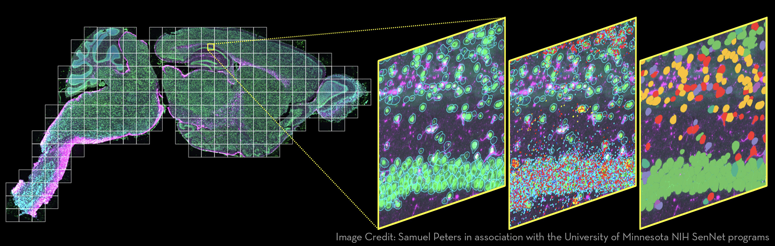
The CosMx Spatial Molecular Imager (SMI) is an integrated system with mature cyclic fluorescent in situ hybridization (FISH) chemistry, high-resolution imaging readout, and interactive data analysis and visualization software. This high-plex in situ analysis platform provides spatial multiomics with formalin-fixed paraffin-embedded (FFPE) and fresh frozen (FF) tissue samples at cellular and subcellular resolution, and enables rapid quantification and visualization of up to 6,000 RNA and 64 validated protein analyte
Key Features:
- Multi-modal cell segmentation process provides accurate cell boundaries detection: CosMx cell segmentation uses cell membrane and morphology marker protein images, machine-learning augmented cell segmentation algorithm and transcript-based segmentation refinement to achieve precise single-cell segmentation in morphologically intact tissue.
- High-Plex Single-Cell Spatial: RNA assays enable high-plex analysis of up to 6000 genes at cellular and sub-cellular levels. RNA assays are designed to provide robust cell typing, cell-cell interaction analysis, and more in a wide range of human and mouse tissues and disease states. Assays are designed to provide a wealth of genomic breadth with ultra-high-plex RNA panels that can provide high numbers of genes detected per sample. Profile expression of highly curated targets at subcellular resolution and customize some assays with your own targets. Panels: Human Whole Transcriptome, Human 6K Discovery, Universal Cell Characterization, Mouse Neuroscience, Human Immuno-Oncology.
- High Resolution Spatial Proteomic Analysis: Protein assays enable high-plex analysis of up to 64 proteins with spatial context at single-cell resolution. Protein assays provide unbiased spatial proteomic analysis of tissue sections to expand the number of markers you can profile compared to traditional immunohistochemical and immunofluorescent methods. All protein assays are validated for use in multiplex. Panels: Mouse Neuroscience, Human Immuno-Oncology.
- Analyze and visualize CosMx spatial data with flexibility: From AtoMx, the UIC exports CosMx data (counts matrices, cell metadata, decoded transcript information, and image files) in formats readable by a variety of open-source tools, allowing researchers to develop fully custom analysis pipelines using their own preferred workflows. CosMx target decoding and segmentation are completed in the AtoMx Spatial Informatics Platform. Here, researchers can visualize spatial data using image overlays of morphology markers, decoded transcripts, and segmentation masks.
Additional Information:
Sample Requirements
- Sample must fit in specified region on slide (20mm x 15mm)
Relevant Publications
Wang, Huan & Huang, Ruixu & Nelson, Jack & Gao, Ce & Trans, Miles & Yeaton, Anna & Felt, Kristen & Pfaff, Kathleen & Bowman, Teri & Rodig, Scott & Wei, Kevin & Goods, Brittany & Farhi, Samouil. (2023). Systematic benchmarking of imaging spatial transcriptomics platforms in FFPE tissues. bioRxiv : the preprint server for biology. 10.1101/2023.12.07.570603.