GeoMx Digital Spatial Profiler (DSP)
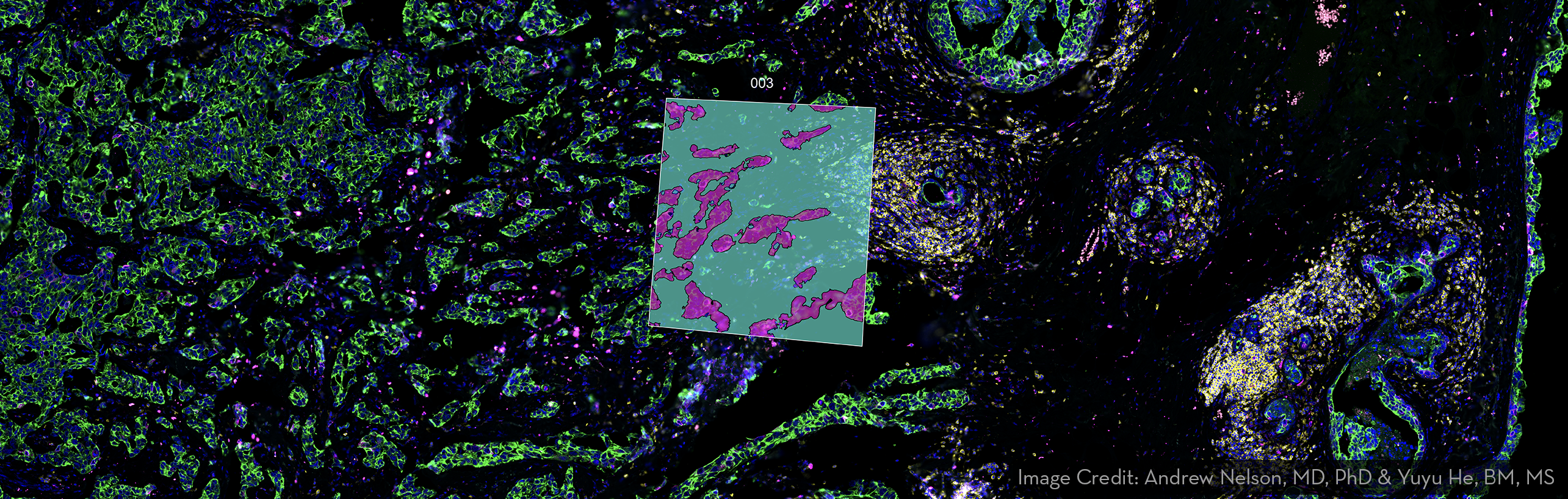
The GeoMx Digital Spatial Profiler (DSP) combines standard immunofluorescence techniques with digital optical barcoding technology to perform highly multiplexed, spatially resolved profiling experiments. In a single reaction, the DSP technology performs whole slide imaging with up to four fluorescent stains to capture tissue morphology and select regions of interest enabling spatially resolved high plex profiling of RNA and protein targets on FFPE and frozen samples. Antibody or nucleic acid probes are coupled to photocleavable oligonucleotide tags and are hybridized to targets in slide mounted tissue sections. Then, oligonucleotide tags are released from discrete regions of the tissue via UV exposure. Released tags are quantified by nCounter technology or Illumina Next Generation Sequencing (NGS). Counts are mapped back to tissue location, yielding a spatially resolved digital profile of analyte abundance.
Key Features:
- Quickly resolve tissue heterogeneity and the complexity of microenvironments: The GeoMx Digital Spatial Profiler (DSP) is a highly flexible and robust spatial multiomic platform for analysis of FFPE and fresh frozen tissue sections. GeoMx is the only spatial biology platform that non-destructively profiles expression of RNA and protein from distinct tissue compartments and cell populations with an automated and scalable workflow that integrates with standard histology staining.
- GeoMx RNA assays allow you to profile the whole transcriptome: GeoMx RNA Assays are pre-validated and modular to provide flexibility and support a range of research needs. Available content covers immunology, immuno-oncology, neurodegeneration, and neuroinflammation with a rapidly growing pipeline.
- High-plex Spatial Proteomics: GeoMx Protein Assays enable quantitative, spatial analysis of up to 570+ proteins, vastly expanding the number of markers you can profile from a single tissue section compared to traditional immunohistochemical methods. All assays are validated for use in multiplex.
- Stain whole slides with assay probes and up to four fluorescent markers and then image them on the instrument: Choose which regions to profile and segment each region using qualified and verified morphology markers, decide which regions of the tissue you want to profile, and using masking, optionally profile two or more tissue compartments or cell types in each region. Quantify RNA and protein expression levels with direct, digital counting of barcodes using the UMGC nCounter or Illumina sequencing platforms.
Additional Information:
Sample Requirements
- 100-150 cells are needed at minimum
- Samples must fit in specified region on slide (35.4mm x 14.1mm)